
 The closely appressed, thin, upper and lower wing membranes are supported by a system of tubular veins whose main function is to strengthen the wings. The venation pattern is referred to by a specialised notation and the patterning of the veins is of great value in insect classification. Although other systems were used previously, primarily numerical ones, the standard nomenclature used today is the Comstock-Needham system (Comstock & Needham, 1898-1899; Comstock, 1918; Lameere, 1922). In this website, where original descriptions involve other systems of wing venation nomenclature, these have been translated to the Comstock terminology as necessary. In the primitive lepidopteran families the wing venation is similar on fore and hindwings (homoneurous). The pyraloid and thyridoid venation is of the heteroneurous type where the pattern is different on the FW and HW, with a reduced number of HW veins.
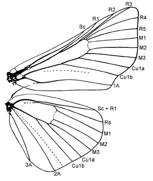
The veins divide the wing into spaces or cells. In the Comstock-Needham system the terminology of the cells is derived from veins which form their anterior margins. The most obvious cell between the stem of Cu1A and the stem of R1 is the discal cell or simply ‘the cell’. The distal end of this is usually ‘closed’ by a series of cross veins between M1, M2 and M3 and the fused stem of R4 and R5 (R4+5) but in a few subfamilies this may be partly ‘open’ lacking the cross veins.